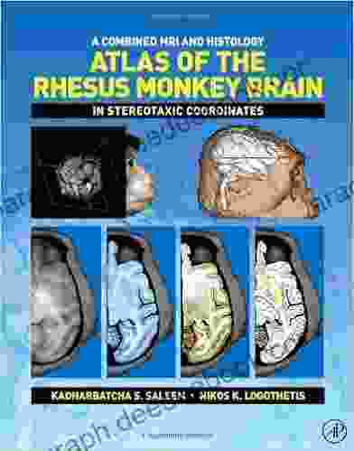
A Comprehensive Guide to the Combined MRI and Histology Atlas of the Rhesus Monkey Brain in Stereotaxic Coordinates

The rhesus monkey (Macaca mulatta) is a widely used animal model in biomedical research, particularly in neuroscience. The development of high-resolution magnetic resonance imaging (MRI) and histology techniques has enabled the creation of detailed anatomical atlases of the rhesus monkey brain. These atlases provide researchers with a valuable tool for studying the brain structure and function in both normal and disease states.
The Combined MRI and Histology Atlas of the Rhesus Monkey Brain in Stereotaxic Coordinates provides a comprehensive overview of the brain's anatomy, with high-resolution MRI images and corresponding histological sections. The atlas is divided into six volumes, each covering a different region of the brain:
5 out of 5
| Language | : | English |
| File size | : | 11761 KB |
| Text-to-Speech | : | Enabled |
| Print length | : | 336 pages |
* Volume 1: Cerebral Cortex * Volume 2: Hippocampus and Amygdala * Volume 3: Thalamus and Basal Ganglia * Volume 4: Cerebellum and Brainstem * Volume 5: Spinal Cord * Volume 6: Cranial Nerves
Each volume contains a series of coronal, sagittal, and axial MRI images, along with corresponding histological sections stained for different cellular markers. The histological sections provide detailed information about the cytoarchitecture of the brain, while the MRI images provide a three-dimensional view of the brain's structure.
The atlas is a valuable resource for researchers in a variety of disciplines, including neuroscience, anatomy, and neurology. It can be used for a variety of purposes, such as:
* Studying the brain structure and function in normal and disease states * Planning and conducting experiments * Teaching neuroanatomy * Developing new imaging techniques
MRI and Histology Techniques
The MRI images in the atlas were acquired using a 7T MRI scanner, which provides high-resolution images with excellent contrast between different brain tissues. The histological sections were stained using a variety of techniques, including hematoxylin and eosin (H&E),Nissl, and immunohistochemistry.
H&E staining is a basic histological technique that stains cell nuclei blue and cytoplasm pink. Nissl staining is a more specific histological technique that stains neurons blue. Immunohistochemistry is a technique that uses antibodies to stain specific proteins, such as neurotransmitters or receptors.
Atlas Organization
The atlas is organized into six volumes, each covering a different region of the brain. Each volume contains a series of coronal, sagittal, and axial MRI images, along with corresponding histological sections. The MRI images are labeled with the corresponding stereotaxic coordinates, which allows researchers to easily identify the location of a particular brain region.
The histological sections are stained for different cellular markers, such as Nissl, myelin, and immunohistochemistry. The Nissl-stained sections provide detailed information about the cytoarchitecture of the brain, while the myelin-stained sections provide information about the white matter tracts. The immunohistochemistry-stained sections provide information about the distribution of specific proteins, such as neurotransmitters or receptors.
Applications
The Combined MRI and Histology Atlas of the Rhesus Monkey Brain in Stereotaxic Coordinates has a wide range of applications in neuroscience research. It can be used for a variety of purposes, such as:
* Studying the brain structure and function in normal and disease states * Planning and conducting experiments * Teaching neuroanatomy * Developing new imaging techniques
The atlas is a valuable resource for researchers in a variety of disciplines, including neuroscience, anatomy, and neurology. It provides a comprehensive overview of the rhesus monkey brain's anatomy, with high-resolution MRI images and corresponding histological sections.
The Combined MRI and Histology Atlas of the Rhesus Monkey Brain in Stereotaxic Coordinates is a valuable resource for researchers in neuroscience. It provides a comprehensive overview of the brain's anatomy, with high-resolution MRI images and corresponding histological sections. The atlas can be used for a variety of purposes, such as studying the brain structure and function in normal and disease states, planning and conducting experiments, teaching neuroanatomy, and developing new imaging techniques.
5 out of 5
| Language | : | English |
| File size | : | 11761 KB |
| Text-to-Speech | : | Enabled |
| Print length | : | 336 pages |
Do you want to contribute by writing guest posts on this blog?
Please contact us and send us a resume of previous articles that you have written.
 Novel
Novel Chapter
 Reader
 Library
 E-book
 Magazine
 Newspaper
 Sentence
 Bookmark
 Shelf
Shelf Glossary
 Bibliography
 Foreword
 Preface
 Synopsis
 Footnote
 Scroll
 Tome
Tome Classics
 Biography
 Autobiography
 Memoir
 Dictionary
 Narrator
 Character
 Resolution
 Catalog
 Card Catalog
 Borrowing
 Stacks
 Periodicals
 Study
Study Academic
 Journals
 Rare Books
 Special Collections
 Study Group
 Storytelling
 Reading List
 Textbooks
 Enjoy Discovering
 E E Cummings
 Daniel Ankele
 Robert Gaylon Ross
 Diana Gabaldon
 Christopher Baugh
 Joseph C Zinker
 Michael Slack
 Steven E Siry
 Gordon Brown
 Ioannis Anastassakis
 Rajan Suri
 Jan Sandford
 Mark Pendergrast
 Johnny Marr
 Antony Loewenstein
 Dorothy Eden
 Martin Turnbull
 Wilfredo Gonzalez
 Oliver Clarke
Light bulbAdvertise smarter! Our strategic ad space ensures maximum exposure. Reserve your spot today!

 Brandon CoxBeginner's Guide to Value Investing: The Proven Trading Strategies to Retire...

 Oliver FosterThe Society of the Spectacle: A Critical Examination of Mass Media and Its...
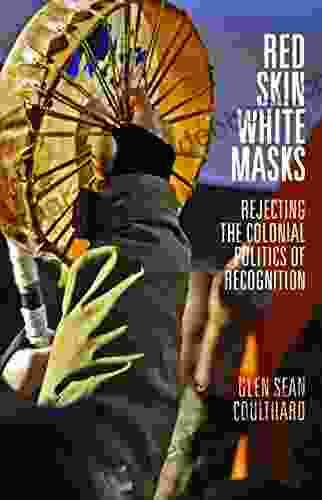
 Langston HughesUnveiling the Significance of Red Skin, White Masks: A Journey into Identity,...
 Miguel NelsonFollow ·11.7k
 Douglas AdamsFollow ·14.3k
 Oscar WildeFollow ·18.4k
 Gary ReedFollow ·2k
 Ken SimmonsFollow ·17.1k
 Steve CarterFollow ·12k
 Dan BrownFollow ·15.8k
 Theodore MitchellFollow ·13.7k

 Ricky Bell
The Marriage: An Absolutely Jaw-Dropping Psychological...
In the realm of...

 Ray Blair
Discover the Enchanting Charm of Budapest and Its...
Nestled in the heart of...

 Tyrone Powell
Huddle: How Women Unlock Their Collective Power
Huddle is a global movement that empowers...

 Grayson Bell
The Coin Story of the Holocaust: A Symbol of Hope and...
In the depths of the...

 Virginia Woolf
Folklore Performance and Identity in Cuzco, Peru: A...
Nestled amidst...

 Dylan Mitchell
The Enduring Love Story of Héloïse and Abélard: A Tale of...
An Intellectual Passion In the heart of...
5 out of 5
| Language | : | English |
| File size | : | 11761 KB |
| Text-to-Speech | : | Enabled |
| Print length | : | 336 pages |


