
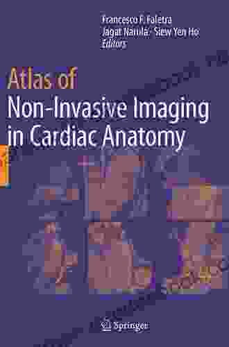
Atlas of Non Invasive Imaging in Cardiac Anatomy

The Atlas of Non Invasive Imaging in Cardiac Anatomy is a comprehensive guide to the use of non invasive imaging techniques in the study of cardiac anatomy. The atlas provides detailed descriptions of the various imaging modalities, including echocardiography, computed tomography (CT),and magnetic resonance imaging (MRI),and their applications in the diagnosis and management of cardiac disease.
5 out of 5
| Language | : | English |
| File size | : | 87776 KB |
| Text-to-Speech | : | Enabled |
| Screen Reader | : | Supported |
| Enhanced typesetting | : | Enabled |
| Print length | : | 272 pages |
The atlas is a valuable resource for cardiologists, radiologists, and other healthcare professionals who use non invasive imaging techniques in their practice. The atlas can also be used by students and researchers who are interested in learning more about the use of non invasive imaging in cardiac anatomy.
History and Development
The development of non invasive imaging techniques in cardiac anatomy began in the early 20th century with the of X-ray fluoroscopy. Fluoroscopy allowed physicians to visualize the heart and its major blood vessels in real time, and it was quickly adopted for use in the diagnosis and management of cardiac disease.
In the 1950s, echocardiography was developed as a non invasive method for visualizing the heart and its structures. Echocardiography uses sound waves to create images of the heart, and it is now one of the most widely used imaging modalities in cardiac anatomy.
In the 1970s, CT and MRI were developed as non invasive imaging techniques for visualizing the heart and its structures. CT uses X-rays to create cross-sectional images of the heart, while MRI uses magnetic fields and radio waves to create images of the heart.
The development of non invasive imaging techniques in cardiac anatomy has revolutionized the way that we diagnose and manage cardiac disease. These techniques allow us to visualize the heart and its structures in great detail, and they have led to the development of new treatments for cardiac disease.
Applications
Non invasive imaging techniques are used in a wide variety of applications in cardiac anatomy. These applications include:
*
*
*
*
Non invasive imaging techniques are also used in research to study the structure and function of the heart.
Future Prospects
The future of non invasive imaging in cardiac anatomy is bright. New imaging techniques are being developed all the time, and these techniques are providing us with increasingly detailed images of the heart and its structures.
One of the most promising new imaging techniques is cardiac magnetic resonance imaging (CMR). CMR uses magnetic fields and radio waves to create images of the heart, and it provides excellent visualization of the heart's anatomy and function. CMR is also being used to develop new treatments for cardiac disease.
Another promising new imaging technique is cardiac computed tomography (CCT). CCT uses X-rays to create cross-sectional images of the heart, and it provides excellent visualization of the heart's arteries and veins. CCT is also being used to develop new treatments for cardiac disease.
The development of new non invasive imaging techniques in cardiac anatomy is leading to new ways to diagnose, manage, and treat cardiac disease. These techniques are providing us with a better understanding of the heart and its function, and they are helping us to improve the lives of people with cardiac disease.
The Atlas of Non Invasive Imaging in Cardiac Anatomy is a comprehensive guide to the use of non invasive imaging techniques in the study of cardiac anatomy. The atlas is a valuable resource for cardiologists, radiologists, and other healthcare professionals who use non invasive imaging techniques in their practice. The atlas can also be used by students and researchers who are interested in learning more about the use of non invasive imaging in cardiac anatomy.
The future of non invasive imaging in cardiac anatomy is bright. New imaging techniques are being developed all the time, and these techniques are providing us with increasingly detailed images of the heart and its structures. These techniques are leading to new ways to diagnose, manage, and treat cardiac disease, and they are helping us to improve the lives of people with cardiac disease.
5 out of 5
| Language | : | English |
| File size | : | 87776 KB |
| Text-to-Speech | : | Enabled |
| Screen Reader | : | Supported |
| Enhanced typesetting | : | Enabled |
| Print length | : | 272 pages |
Do you want to contribute by writing guest posts on this blog?
Please contact us and send us a resume of previous articles that you have written.
 Novel
Novel Page
Page Chapter
 Text
Text Genre
Genre Paperback
 E-book
 Paragraph
 Glossary
 Preface
 Annotation
 Manuscript
 Scroll
 Codex
Codex Tome
Tome Bestseller
 Library card
 Narrative
 Biography
 Memoir
 Reference
 Encyclopedia
 Thesaurus
 Narrator
 Character
 Resolution
 Card Catalog
 Borrowing
 Stacks
 Archives
 Scholarly
 Journals
 Reading Room
 Rare Books
 Thesis
 Dissertation
 Awards
 Book Club
 Theory
 Textbooks
 Kenney Jones
 Suzanne Clothier
 Margaret Byron
 Jared Sparks
 Kaye Draper
 Crystal Kinn Tarver
 Will Adams
 Christine Haynes
 C A J Coady
 Stephen J King
 Richard L Collins
 Pat Walsh
 Joanna Cazden
 Jim Clark R P T
 Jace Weaver
 Jill Mcdonald
 Hindol Sengupta
 Leah Marie Brown
 Jg Jones
 Bryan Mccann
Light bulbAdvertise smarter! Our strategic ad space ensures maximum exposure. Reserve your spot today!


 Ralph EllisonEvery Zombie Eats Somebody Sometime: An In-Depth Exploration of the Undead in...
 Kurt VonnegutFollow ·6.7k
 Sam CarterFollow ·2.2k
 Jayden CoxFollow ·12.5k
 Chuck MitchellFollow ·3.2k
 Jacob FosterFollow ·3.4k
 Edmund HayesFollow ·7.8k
 George R.R. MartinFollow ·12.3k
 Haruki MurakamiFollow ·16.4k

 Ricky Bell
The Marriage: An Absolutely Jaw-Dropping Psychological...
In the realm of...

 Ray Blair
Discover the Enchanting Charm of Budapest and Its...
Nestled in the heart of...

 Tyrone Powell
Huddle: How Women Unlock Their Collective Power
Huddle is a global movement that empowers...

 Grayson Bell
The Coin Story of the Holocaust: A Symbol of Hope and...
In the depths of the...
 Virginia Woolf
Folklore Performance and Identity in Cuzco, Peru: A...
Nestled amidst...

 Dylan Mitchell
The Enduring Love Story of Héloïse and Abélard: A Tale of...
An Intellectual Passion In the heart of...
5 out of 5
| Language | : | English |
| File size | : | 87776 KB |
| Text-to-Speech | : | Enabled |
| Screen Reader | : | Supported |
| Enhanced typesetting | : | Enabled |
| Print length | : | 272 pages |
